
 Türkçe
 Deutsch
 Français
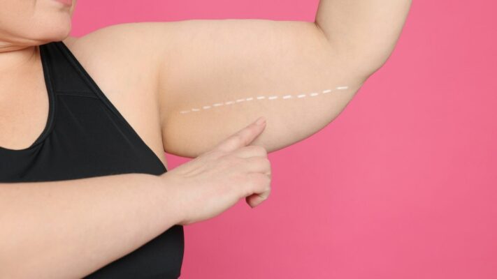
Plastic surgery is the art of reshaping and repairing various parts of the body. This surgical discipline focuses both on resolving functional problems and improving aesthetic appearance. Its primary goal is to restore bodily integrity and improve the person’s quality of life, whether the issue involves tissue loss caused by an accident, a congenital difference, or changes that develop over time. Advanced surgical techniques are used to restore body structures to their most ideal form and function.












EFC Surgical Medical Center
Comprehensive Care: From Initial Consultation to Follow-Up.
EFC Surgical Medical Center is a center of excellence specializing in the most meticulous fields of surgical medicine, from aesthetic surgery to interventional treatments—where every step progresses with refined attention. Medical excellence, aesthetic precision, and uncompromising ethical standards converge on the same path. Our subspecialty-trained experts aim to achieve natural and reliable results by delivering evidence-based care supported by modern imaging, standardized protocols, and safety systems. From consultation to recovery, your care is coordinated end-to-end with clear communication, transparent planning, and genuine respect for your health.
Are the Fields of Plastic Surgery Limited Only to Aesthetics?
Although aesthetic or cosmetic surgery is the first thing that comes to mind in society when plastic surgery is mentioned, it is actually a much broader umbrella. Plastic surgery essentially covers two main subspecialties: Reconstructive surgery and Cosmetic surgery.
What Is Reconstructive Surgery?
By definition, reconstructive surgery focuses on structures of the body that are “abnormal” or have lost their function. The primary clinical objective is to restore normal function and appearance by correcting congenital defects, injuries caused by accidents, infections, or diseases such as cancer.
Some common examples of reconstructive surgery include:
- Cleft lip and palate repair
- Breast repair following breast cancer (reconstruction)
- Correction of contractures that develop after severe burns
- Reattachment of limbs severed by trauma (replantation)
- Repairing tissue loss after tumor removal
From clinical and administrative perspectives, reconstructive surgery is generally considered “medically necessary” and is therefore assessed within the scope of health insurance coverage.
What Is Cosmetic (Aesthetic) Surgery?
Unlike reconstructive surgery, cosmetic surgery is performed to reshape or alter the body’s “normal” structures. The purpose of cosmetic surgery is entirely aesthetic; its sole objective is to improve the patient’s existing appearance and change their form.
Some common cosmetic procedures include:
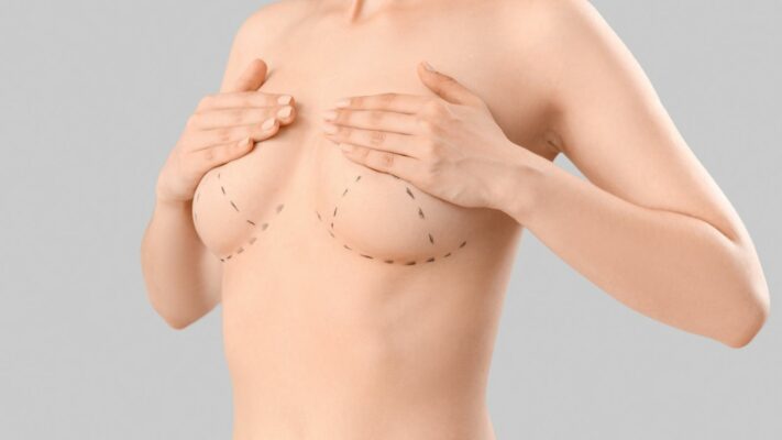
- Breast augmentation or reduction
- Liposuction (Fat removal)
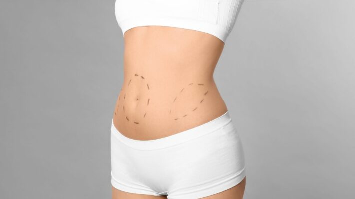
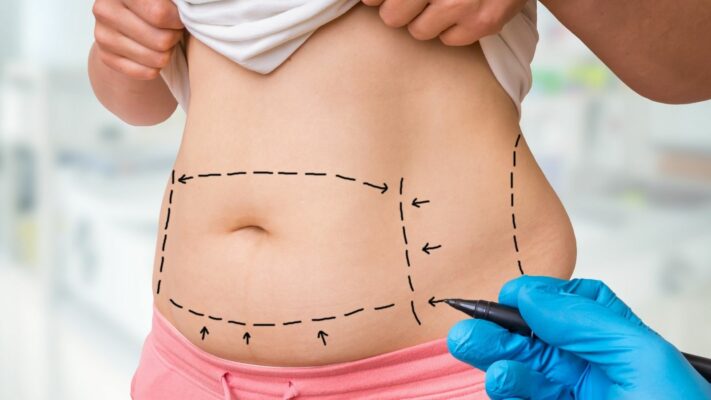
- Abdominoplasty (Tummy tuck)
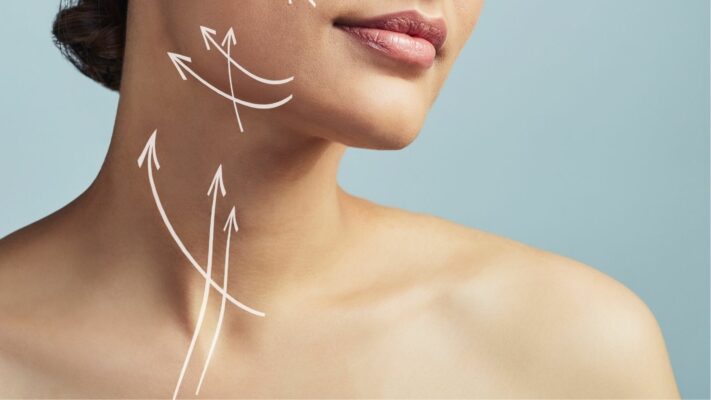
- Rhytidectomy (Facelift)
- Rhinoplasty (Nose surgery)
Because cosmetic surgery is not considered medically necessary, it is classified as an “optional” (elective) procedure and is generally not covered by health insurance.
Is the Boundary Between Aesthetics and Reconstruction in Plastic Surgery Always Clear?
Combining these two fields is the fundamental identity of plastic surgery. In practice, however, the distinction between “reconstructive” and “cosmetic” is often unclear. This uncertainty may create difficulties in terms of both clinical classification and insurance reimbursement.
Rhinoplasty (nose surgery) is the most classic example of this uncertainty.
A rhinoplasty performed solely to “improve the appearance of the nose” is classified as “cosmetic.” However, the exact same surgical procedure required to “restore normal breathing and normal appearance after a severe nasal fracture” is classified as “reconstructive.”
This demonstrates that the classification depends not on the surgical technique itself but on the cause of the defect, such as congenital or traumatic causes versus normal anatomy, and the primary purpose, such as restoring function versus improving appearance.
Who Is the Right Candidate for Aesthetic Surgery?
Aesthetic surgery is performed to improve a patient’s “appearance and self-confidence.” The primary motivation of patients seeking aesthetic procedures is generally dissatisfaction with their body image. Therefore, patient selection requires not only a physical assessment but also an evaluation of the patient’s psychological condition and motivations.
What Is the Role of Psychological Evaluation?
A critical component of consultation before an aesthetic procedure is a comprehensive psychological evaluation. The primary purpose of this assessment is to identify “patient types” or forms of psychopathology that may constitute an obstacle to surgical intervention.
One of the primary conditions of concern is Body Dysmorphic Disorder (BDD). Patients with BDD may have unrealistic expectations that cannot be corrected surgically or a distorted perception of their “normal” anatomy. In such cases, these patients may be better treated “by a mental health professional rather than an aesthetic surgeon.” This psychological evaluation is an ethical and clinical responsibility based on the surgeon’s individual judgment.
What Are the Common Complications of Plastic Surgery?
Like all surgical specialties, plastic surgery inherently carries risks.
Like all operations, plastic surgery carries certain risks. The most common potential complications include:
- Surgical site infection
- Hematoma (Blood accumulation)
- Seroma (Fluid accumulation)
- Separation of wound edges
- Tissue death (necrosis)
- Venous thromboembolism (VTE)
Hematomas and seromas are common wound collections that may jeopardize surgical outcomes.
Why Is the Risk of Venous Thromboembolism (VTE) So Important?
Venous thromboembolism (VTE), which includes deep vein thrombosis (DVT) and pulmonary embolism (PE – blood clot in the lung), is one of the most serious and potentially life-threatening complications in plastic surgery. The risk is not the same for all procedures; abdominoplasty (tummy tuck), in particular, is described as “one of the plastic surgery procedures with the highest risk of venous thromboembolism.”
Therefore, determining the patient’s risk of blood clots using tools such as the Caprini scoring system is critically important. High-risk patients may require prophylaxis with blood-thinning (anticoagulant) medications.
However, this situation creates a critical clinical conflict, particularly in high-risk aesthetic procedures. Procedures with the highest risk of blood clots, such as tummy tucks, also involve “large raw surface areas and tissue separation (dissection).” Because this extensive dissection significantly increases the risk of hematoma or bleeding, “using blood thinners before surgery” may be risky.
At this point, the surgeon must make a difficult clinical decision: Reducing the risk of blood clots with blood thinners may directly cause a surgical complication (hematoma), which may jeopardize tissue health or require another operation.
Contact us now to get detailed information about our treatments and procedures and to schedule an appointment!
How Is Facelift (Rhytidectomy) Surgery Performed?
The facelift procedure has undergone significant evolution over the years. Early techniques in the early 1900s were based solely on cutting away excess skin. However, these methods based only on tightening the skin lost their effectiveness over time due to the “natural elasticity of the skin.” This limitation prompted surgeons to explore the deeper layers beneath the skin to achieve more durable and natural-looking rejuvenation.
What Is the SMAS Technique?
The pivotal moment in modern facelift surgery was the anatomical description of the SMAS (Superficial Musculoaponeurotic System) in 1976. The SMAS is a distinct fascial (connective tissue) layer located immediately beneath the skin, continuous with the muscles in the neck, and surrounding the facial expression muscles.
In SMAS techniques, this underlying SMAS layer is manipulated after the skin is lifted. There are two main methods: folding the SMAS onto itself (plication) or removing and tightening part of it (SMASectomy). These techniques are primarily used to correct jowls resulting from sagging cheeks and sagging of the neck.
What Is a Deep Plane Facelift?
The further evolution of facelift surgery moved the surgical plane deeper, into the sub-SMAS plane. This is the critical difference in these techniques. It allows the surgeon to lift the SMAS, the overlying cheek fat, and the skin as a single integrated unit. Deep plane facelift surgery releases the key retaining ligaments of the face, allowing the midface to be fully lifted and repositioned.
Which Facelift Technique Is Better?
This remains a subject of debate among surgeons. Because the lifting procedure is anchored to deep tissue, deep plane techniques offer the advantage of tension-free skin closure and may be more effective in lifting the midface.
However, these benefits come with increased technical difficulty. Sub-SMAS dissection is more complex, carries “greater risk to the facial nerve,” and generally requires a longer recovery period. While some studies support the superior results of the deep plane technique, other studies have found no significant difference in the long term. This suggests to the practitioner that meticulous application of the selected technique and the surgeon’s experience may be more critical than the specific plane of dissection.
How Is Eyelid Aesthetic Surgery (Blepharoplasty) Performed?
Upper eyelid aesthetic surgery generally involves removing excess skin (dermatochalasis), loosened muscle, and herniated fat pads:
The lower eyelid is more complex, and the approach is determined according to the patient’s primary complaint.
Which Methods Are Used in Lower Eyelid Surgery?
There are two main approaches to lower eyelid surgery, and the choice depends on the patient’s anatomy:
Transcutaneous (External Incision) Approach: The incision is made through the skin immediately below the lash line, approximately 1 mm beneath it. This approach is used primarily for patients with excess lower eyelid skin and skin/muscle laxity. This method allows excess skin to be removed directly but carries a higher risk of postoperative lower eyelid malposition, such as ectropion or outward turning of the eyelid, due to vertical scarring and tension.
Transconjunctival (Internal Incision) Approach: This is an internal approach. The incision is made through the conjunctiva on the inside of the lower eyelid, leaving no external scar. This approach is primarily indicated for patients without significant excess skin, generally younger patients, but who complain of herniated orbital fat pads (“bags”). It leaves no external scar, and the risk of eyelid retraction is minimal.
What Is the Modern Approach to Eyelid Aesthetic Surgery?
The philosophy of modern eyelid surgery has undergone a significant change. “Historically, lower eyelid aesthetic surgery was a reductive procedure focused on removing skin, muscle, and fat.” This aggressive approach often resulted in a “hollow” and “operated” appearance around the eyes.
The “new concept” or “modern eyelid surgery” instead focuses on “preserving periorbital volume.” This is achieved through “fat preservation and repositioning.” In this technique, the herniated orbital fat is not removed; instead, it is advanced and spread over the orbital rim to fill the tear trough. This represents a fundamental shift in understanding that treats aging around the eyes not merely as excess tissue but as volume loss and sagging.
What Are the Open and Closed Approaches in Rhinoplasty Surgery?
This is one of the questions patients ask most frequently and represents a surgical trade-off: Exposure (Visibility) vs. Tissue Preservation.
Open (External) Rhinoplasty Approach
In this technique, in addition to incisions inside the nose, a small incision is made in the skin bridge between the two nostrils (columella). This allows the surgeon to lift the nasal skin completely and directly view the underlying cartilage and bone framework.
The open approach is preferred when the surgeon needs to see the nasal framework. The main reasons include:
- Complex nasal tip deformities
- Extensive alteration of the nasal dorsum
- Severe nasal deviations
- Structural repairs requiring grafts (cartilage patches)
Closed (Endonasal) Rhinoplasty Approach
In this approach, all incisions are made inside the nostrils. There is no visible external scar. The surgeon accesses the nasal framework by working through these internal incisions.
The closed approach is used for less extensive changes and in cases where preserving the soft tissue envelope is important:
- Simpler nasal tip problems
- Minimal intervention on the nasal dorsum
- Patients with thin skin
In exchange for maximum visualization of the surgical framework, the open approach sacrifices an invisible scar by leaving a small scar on the columella. The closed approach preserves the columella but limits visibility. The surgeon’s decision is strategic: Is the complexity of the deformity great enough to require the complete visibility provided by the open approach?
What Is Preservation Rhinoplasty?
Preservation rhinoplasty is a surgical philosophy that can be applied to both open and closed approaches and emphasizes “bone-cartilage preservation.” Instead of aggressively “breaking” and removing the hump on the nasal dorsum, “impaction” techniques, which lower the dorsum by removing underlying supporting tissues, are frequently used to lower the nasal dorsum while preserving its natural structural integrity.
What Is Considered in Breast Augmentation Surgery?
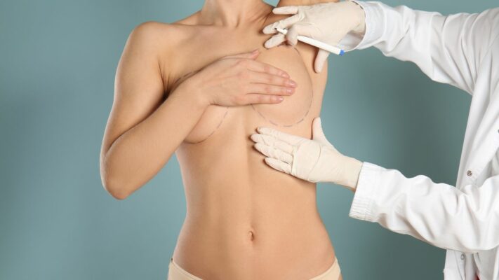
When planning breast augmentation surgery, two fundamental decisions are made: the type of implant (silicone or saline, smooth or textured surface) and where the implant will be placed.
What Is the Implant Placement Plane?
There are two main anatomical pockets in which the implant can be placed:
- Subglandular (Above the muscle): The implant is placed behind the breast gland but in front of the chest muscle (pectoralis major).
- Submuscular (Below the muscle): The implant is placed partially or completely behind the chest muscle.
What Is the Most Common Complication of Breast Augmentation Surgery?
Capsular contracture (CC) is the “most common complication” after implant-based breast surgery. It is defined as an “excessive fibrotic (hardening) foreign-body reaction” by the body to the implant. This process leads to the formation of a thick, fibrous capsule (membrane) surrounding the implant. Over time, this capsule tightens, causing the breast to become hard, painful, and aesthetically deformed.
The “gold standard” treatment for symptomatic (Severe-Grade) CC is surgical intervention (capsulectomy – removal of the capsule). However, even surgical management has a “significant recurrence rate.”
Contact us now to get detailed information about our treatments and procedures and to schedule an appointment!
How Can the Risk of Capsular Contracture Be Reduced?
Evidence indicates that certain factors increase the risk. When these factors occur together, the risk profile rises:
- Subglandular (above the muscle) placement
- Smooth-surfaced implants
- Silicone-filled implants (compared with saline)
This creates a clear decision tree for the surgeon. The highest-risk configuration can be described as a smooth-surfaced silicone implant placed above the muscle. Conversely, the evidence-based configuration for minimizing the risk of CC is a textured-surface implant placed below the muscle.
How Are Breast Lift (Mastopexy) and Reduction Surgery Planned?
When planning a mastopexy (lift) or reduction surgery, the surgeon must resolve two separate anatomical problems simultaneously: (1) reshaping the skin envelope (skin “bra”) to correct sagging and reduce volume, and (2) repositioning the nipple-areola complex (NAC) on a viable vascular stalk (pedicle).
Which Incision Patterns Are Used for the Skin Envelope?
The incision pattern used is selected according to the degree of sagging and the amount of reduction:
- Periareolar (Only around the areola)
- Vertical (‘Lollipop’ incision)
- Wise Pattern (‘Inverted-T’ or ‘anchor’ incision)
The periareolar incision leaves the least scarring but is suitable only for mild sagging. The vertical incision provides good projection for moderate sagging. The Wise pattern leaves the most scarring but provides maximum control in cases of the most severe sagging and large-volume reductions.
How Is Nipple Viability (Pedicle) Preserved?
The “pedicle” is the vital “vascular stalk” consisting of glandular tissue, fat, and blood vessels that is preserved and mobilized to maintain the viability and sensation of the Nipple-Areola Complex (NAC).
Different pedicle (vascular stalk) techniques are used to preserve the blood vessels supplying the nipple. The main techniques include:
- Inferior (Lower) Pedicle
- Superomedial (Upper-inner) Pedicle
- Superior (Upper) Pedicle
For example, a common and reliable procedure is the combination of a “Superomedial pedicle with an Inverted-T (Wise) pattern skin resection.”
How Is Breast Repair (Reconstruction) Performed After Mastectomy?
There are two main options for breast reconstruction after mastectomy (breast removal): implant-based repair or autologous repair using the patient’s own tissue.
What Are the Advantages and Disadvantages of Implant-Based Repair?
This is the most common approach. Its popularity results from its technical simplicity and, most importantly, the “absence of donor-site morbidity.”
However, implants carry long-term risks such as capsular contracture and implant rupture. More importantly, implant-based reconstruction has “higher complication rates in irradiated areas,” making it a less preferred option for patients expected to receive radiotherapy after mastectomy.
What Is Repair Using the Patient’s Own Tissue (Autologous Flap)?
Autologous reconstruction uses the patient’s own tissue, generally from the abdomen, back, or thigh, to create a “natural,” soft, and durable breast mound. It is the preferred method for patients expected to receive radiation therapy.
What Is the Difference Between a TRAM Flap and a DIEP Flap?
This is the best example demonstrating the evolution of autologous repair. Both generally create a new breast using skin and fat from the lower abdomen, but there is a vital difference between them:
- Pedicled TRAM Flap: This is an older technique. A large ellipse of skin and fat taken from the lower abdomen is harvested together with part or all of the underlying rectus abdominis muscle. This entire unit remains attached to its blood supply and is tunneled beneath the skin to reach the chest.
Disadvantage: Its main disadvantage is significant donor-site (abdominal) morbidity. Sacrificing the rectus muscle results in high rates of abdominal wall weakness, bulging, and hernia, reaching up to 16%.
- DIEP Free Flap: This is the preferred method in modern practice and is a “muscle-sparing” technique.
Method: Like the TRAM flap, this flap uses lower abdominal skin and fat. However, the surgeon meticulously separates the small blood vessels (perforators) supplying that skin and fat from within the muscle using microsurgery and leaves the muscle entirely in place. The flap is then transferred to the chest after being detached from its blood supply, and its vessels are reattached (anastomosed) to recipient vessels in the chest under microsurgery.
Advantages: This technique provides the same “natural” tissue as a TRAM flap but minimizes abdominal donor-site morbidity, with a 1% hernia rate, by preserving the entire abdominal muscle.
Who Is Abdominoplasty (Tummy Tuck) Surgery Performed On?
Abdominoplasty is a surgical procedure designed to correct two fundamental anatomical problems: excessive skin laxity and separation of the rectus abdominis muscles (diastasis recti). These conditions generally occur after pregnancy or excessive weight loss.
What Is the Difference Between a Full and Mini Tummy Tuck?
The scope of the operation is determined according to the severity of the problem, and there are two main types:
- Full Abdominoplasty
- Mini Abdominoplasty
A full tummy tuck is necessary for severe sagging and muscle separation (diastasis recti) both below and above the navel. During this procedure, the muscles are generally repaired (plication), and the navel is moved to its new position. A mini tummy tuck is suitable for patients who only have mild laxity below the navel and generally do not have muscle separation. This is a more limited procedure, and the navel is not altered.
What Is Lipoabdominoplasty?
Lipoabdominoplasty represents a modern evolution that combines liposuction with abdominoplasty skin excision. In this technique, traditional extensive undermining beneath the abdominal flap is avoided. Instead, liposuction is used for “lipo-dissection.” This approach preserves Scarpa’s fascia and, most importantly, the abdominal wall perforator vessels that supply blood to the flap. By preserving this intact blood supply and minimizing the “dead space” created by extensive undermining, lipoabdominoplasty has been shown to have a lower complication rate, particularly for seroma (fluid accumulation).
What Are the Differences Between Liposuction Methods?
Liposuction can be performed using different technologies. The main methods include:
- Suction-Assisted Lipoplasty (SAL) – Traditional Method
- Ultrasound-Assisted Lipoplasty (UAL / VASER) – Ultrasound Energy
- Power-Assisted Lipoplasty (PAL) – Mechanical Vibration
SAL involves physically breaking down and suctioning fat through the manual back-and-forth movement of a cannula following tumescent fluid infiltration. PAL uses a handpiece that causes the cannula to vibrate rapidly and mechanically; this reduces surgeon fatigue and accelerates the procedure, particularly in large-volume or firm (fibrous) areas.
VASER (UAL), however, is a “fat pretreatment” device. It uses a specialized probe that emits high-frequency ultrasonic energy to selectively emulsify (liquefy) the fat before it is aspirated.
Why Is “High-Definition” Liposculpture (Abdominal Muscle Aesthetics) Performed with VASER?
The fundamental innovation of UAL/VASER is not merely fat removal but tissue selectivity. Ultrasound energy targets fat cells but “does not damage arterial and venous vessels, lymphatics, or fibrous septa (connective tissue).”
This selectivity is what allows the surgeon to perform “high-definition liposculpture,” such as creating a “six-pack” appearance. Traditional SAL is traumatic; working superficially with an SAL cannula causes serious damage to the vessels and connective structures beneath the skin, resulting in irregularities. By preserving these critical non-fat structures, VASER allows the surgeon to work safely in the “immediate subdermal plane” directly beneath the skin and reveal the contours of the muscles.
What Is the “Reconstructive Ladder”?
The “Reconstructive Ladder” is a fundamental concept for wound closure in plastic surgery. It organizes techniques within a hierarchical framework from the simplest to the most complex.
The steps of this ladder, from the simplest to the most complex, are as follows:
- Secondary Healing (Closure by itself)
- Primary Closure (Sutures)
- Skin Graft (Patch)
- Local Flaps (Adjacent tissue)
- Pedicled Flaps (Stalked tissue)
- Free Flaps (Microsurgery)
However, the principle of the “reconstructive elevator” is generally used in modern practice. This means that a surgeon may skip simpler steps to select a more complex option “if it will optimize the outcome,” such as a free flap for a deep wound on the lower leg. The aim is not the simplest closure but the most appropriate and durable repair.
What Is the Difference Between a Graft (Skin Patch) and a Flap?
These two terms form the basis of reconstructive surgery and are often confused.
- Skin Graft: A skin graft is the transfer of skin, consisting of the epidermis and varying amounts of dermis, that has been completely separated from its blood supply to a recipient wound bed. For the graft to survive (“take”), it must be nourished through diffusion (absorption) from the underlying wound bed and then develop new blood vessels into the area.
- Flap: A flap is a unit of tissue that is moved to cover a defect but brings its own blood supply with it and may contain skin, fat, and muscle. This blood supply either remains connected to its original location through a pedicle or is connected to a new blood supply using microsurgery.
What Are the Types of Skin Grafts?
There are two main types of skin grafts, and there is a critical clinical difference between them:
- Split-Thickness Skin Graft (STSG): It includes the epidermis and part of the dermis. Its advantage is that its “take” rate is very high and the donor site, generally the thigh, can heal on its own. It is used to cover large areas, such as burns.
- Full-Thickness Skin Graft (FTSG): It includes the epidermis and the entire dermis. Its advantage is that it provides much better aesthetic and functional outcomes and, most importantly, causes much less secondary shrinkage (contraction). Its disadvantage is that it is more difficult to “take” because it is thicker, and the donor site must be closed with sutures, which limits the size of the graft that can be harvested.
The clinical choice is a trade-off: STSG provides reliable closure, while FTSG provides high-quality functional/aesthetic repair, particularly on the face, hands, and joints.
Blog Posts

Hair Transplant Prices
Hair transplant prices vary depending on the technique to be used, the number of grafts, [...]
Read More
21
Jan
Jan
Most Preferred Countries for Plastic Surgery
Among the most preferred countries for plastic surgery, Turkey, South Korea, the United States, and [...]
Read More
21
Jan
Jan
Plastic Surgery Prices
Plastic surgery prices vary depending on the type of procedure, geographical location, the surgeon’s experience, [...]
Read More
21
Jan
Jan

Physical Therapy Prices
Physical therapy prices vary depending on the number of sessions to be applied, the treatment [...]
Read More
21
Jan
Jan

Dental Treatment Prices
Dental treatment prices vary depending on the type of procedure to be performed, the quality [...]
Read More
21
Jan
Jan
Most Preferred Countries for Dental Treatments
Among the most preferred countries for dental treatments are Turkey, Hungary, Mexico, and Thailand. These [...]
Read More
21
Jan
Jan
Hair Transplant FUE and DHI Techniques – Complete Guide 2026 | EFC Clinic
Everything you need to know about FUE and DHI hair transplant techniques, procedure steps, recovery [...]
Read MoreHair Transplant FUE and DHI Techniques – Complete Guide 2026 | EFC Clinic
Everything you need to know about FUE and DHI hair transplant techniques, procedure steps, recovery [...]
Read More